 Nanooptics Group - Faculty for Chemistry and Pharmacy
Nanooptics Group - Faculty for Chemistry and Pharmacy
Nanooptics Group - Faculty for Chemistry and Pharmacy
We utilize two different scanning near-feld optical microscopic (SNOM) techniques: 1. Tip-enhanced near-field optical microscopy in which the different spectroscopic signatures such as the sample's fluorescence or Raman spectra are detected and 2. Scattering SNOM which probes the sample's elastic scattering response.
1. Tip-enhanced near-field optical microscopy (TENOM)
This technique we use relies on the locally enhanced optical fields close to a laser illuminated, sharp metal tip which acts as an optical nanoantenna. The sample is raster scanned below the tip which can act as a highly confined excitation source providing a considerable signal enhancement and spectroscopic images at very high spatial resolution. Our experimental setup is based on an inverted optical microscope with an x, y scan stage for raster scanning a transparent sample. The excitation can be provided by a variety of laser sources. The laser light is reflected by a dichroic beam splitter and focused by a high numerical aperture objective (1.49 NA) on the sample surface. A sharp gold tip is positioned in the focus of the beam and maintained above the sample surface at a distance of ~1-2 nm by means of a sensitive shear-force feedback mechanism. The gold tips with a radius of 10-15 nm are produced by electrochemical etching. Both Raman scattered light and luminescence are collected with the same objective, transmitted by the beam splitter and separated by a long pass filter. The signal is then detected either by a combination of a spectrograph and a charged coupled device (CCD) or by avalanche photodiodes (APDs). Alternatively, the tip can also be used to enhance the photocurrent or electroluminescence response of the sample. Tip-enhanced near-field microscopy allows for optical imaging with a spatial resolution below 20 nm and spectroscopy on a nanometer scale.
Example: TENOM on CdSe nanowires
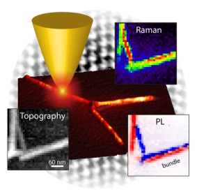 Tip-enhanced photoluminescence (PL) and Raman scattering images of single CdSe nanowires were obtained with a spatial resolution of less than 20 nm. They show that the optical properties of the CdSe NWs vary significantly within a few nanometers leading to strong spatial fluctuations in both PL intensities and energies. Energy gradients of up to 1 meV/nm could be observed. In addition, we showed that two bundled nanowires featuring different band gaps due to different diameters could be spatially resolved by their optical properties.
Example: Visualizing the Local Optical Response of Semiconducting Carbon Nanotubes to DNA-Wrapping
We studied the local optical response of semiconducting single-walled carbon nanotubes to wrapping by DNA segments using high resolution tip-enhanced near-field microscopy. Photoluminescence (PL) near-field images of single nanotubes reveal large DNA-wrapping induced redshifts of the exciton energy that are two times higher than indicated by spatially averaging confocal microscopy. Near-field PL spectra taken along nanotubes feature two distinct PL bands resulting from DNA-wrapped and un-wrapped nanotube segments. The transition between the two energy levels occurs on a length scale smaller than our spatial resolution of about 15 nm. H. Qian et al. Nano Lett. 8, 2706 (2008)
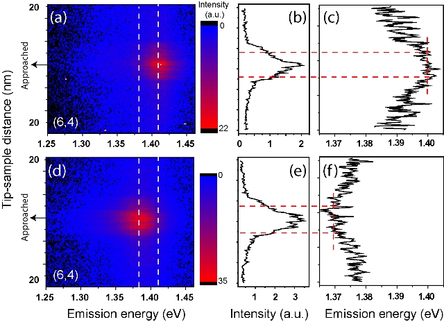
2D photoluminescence maps taken at two different positions of a single (6,4) nanotube (a,d) as a function of tip sample distance. The difference between the two emission energies, marked by two white dashed lines, is due to DNA-wrapping.
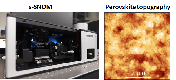
2. Scattering scanning near-field optical microscopy (s-SNOM):
s-SNOM is a scanning probe technique based on atomic force microscopy (AFM), combining an infrared image together with topography. An illuminating infrared (IR) beam is focused onto the probing tip which acts as an antenna, concentrating the incident light at the apex of the tip. Near-field interaction between the tip and the sample is formed by the scattering from the tip. Thus, near field image is created by recording scattered light while scanning the sample surface. What makes s-SNOM is unique is that the spatial resolution is not dependent on the illumination wavelength, but only on the size of the apex, allowing us to construct AFM topography and IR response of the sample surface with nanoscale spatial resolution (~20 nm) at locations of interest. Therefore, nano-FTIR offers nanoscale chemical identification of any substance exhibiting IR vibrational resonances at ultrahigh spatial resolution.