Research topics
General mission
The Müller-Caspary Group understands Microscopy, especially transmission electron microscopy (TEM), as an interdisciplinary challenge. Nowadays, problems in Chemistry, Nanotechnology, Physics, and especially Life Sciences require an understanding of advanced systems at the atomic scale, and even below. Since this demands operating, understanding and modelling TEM imaging at the very performance limits of contemporary Transmission Electron Microscopes, one of the topical foci of the team is fundamental science. This involves the improvement of scattering theories, application of density functional theory and molecular dynamics methods, artificial intelligence and method development in the field of phase retrieval techniques.
With this methodological portfolio, we then gain deep insights into nanotechnological systems such as semiconductor devices or 2D materials, as well as soft and light matter such as proteins and viruses. Besides most of the traditional and established methods in the field of electron and X-ray spectroscopy in a TEM, a key outcome of our recent developments falls in the field of multidimensional electron microscopy. In summary, our activities fall into the following categories:
- Multidimensional electron microscopy
- Structure & chemistry in solid-state nanostructures & 2D materials
- Electrical characterization
- High-contrast low-dose imaging of soft and biological matter
- Development and application of new imaging techniques
- Simulation of electron scattering, Density functional theory, Artificial Intelligence
Multidimensional TEM
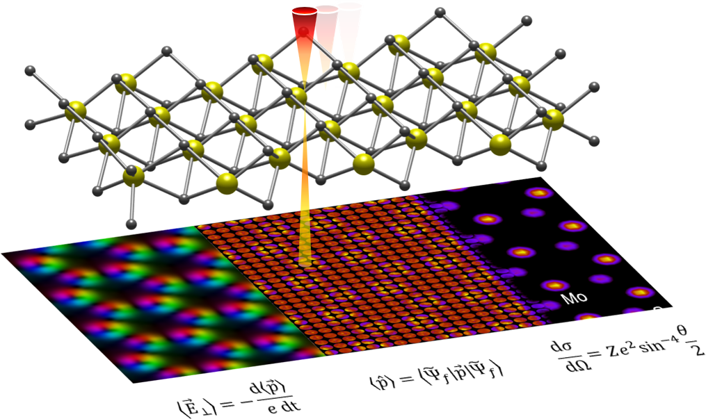
Usually, one can either use a microscope in in imaging mode, or in diffraction mode. In the imaging mode, magnified real-space features of the specimen can be recorded, which is surely the most prevailing driving force in the development of microscopy since its origins centuries ago, and now brought to subatomic resolution with TEM. In diffraction mode, one gets information about periodicities and structure of the specimen, e.g., by exploring positions and intensities of Bragg reflections of crystalline specimens.
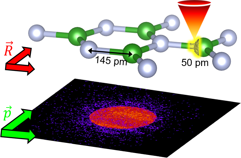
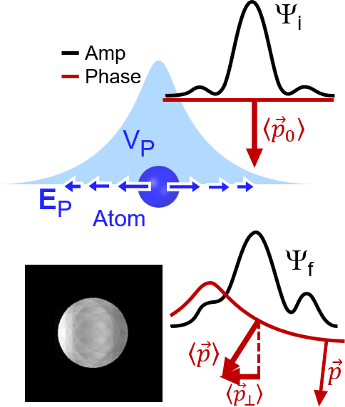
By multidimensional TEM, we aim at getting real- and diffraction space information at ultimate sampling and resolution simultaneously. This is achieved by operating the microscope in diffraction mode, and recording a full diffraction pattern for each position of a focused probe scanning over the specimen point by point in real space. Because the focusing optics allows for probe diameters well below 100pm, we are able to obtain diffraction information from regions being smaller than an atom.
Our microscope facility is equipped with one of the fastest cameras for that purpose. This is necessary because the raster process of the scanning probe needs very fast to cover, e.g., several 100 thousand probe positions. In that respect, we operate a Medpix3 camera in the diffraction plane which can record up to approximately 6000 images per second.
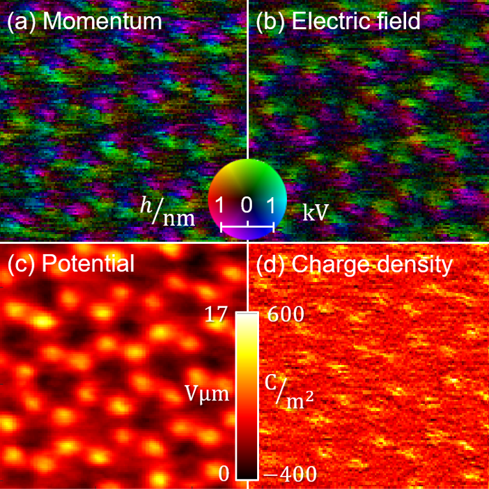
Mapping subatomic electric fields
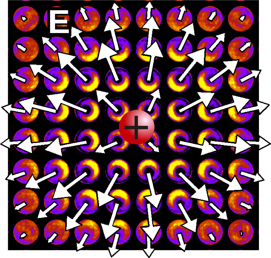
This means that the electrons probe the electric field distribution inside atoms. Our group utilises the resulting diffraction information to measure electric fields, charge densities and electrostatic potentials down to subatomic scales. In very thin materials, such as 2D transition metal dichalcogenides, this is possible with high accuracy.